

Introduction
Potatoes are among the most important economic vegetable crops in all countries due to their high nutritional value [1]. They are the fourth most important economic crop after wheat, corn, and rice [2, 3]. The economic importance of the potato crop in Iraq has increased due to the expansion of cultivated areas [4]. Potatoes are known for their nutritional content of proteins, carbohydrates, vitamins, fibers, fats, and minerals such as phosphorus, potassium, iron, and calcium [5].
Potatoes are susceptible to various diseases [6], including the soil fungus Rhizoctonia solani, which causes black scurf disease. This fungus is globally distributed and poses a real problem in all potato cultivation areas worldwide. The wide familial range of the fungus, its resistance to unfavorable environmental conditions, and its presence in soil and plant residues contribute to its high risk. The fungus enters the plant through natural openings and wounds [7]. The fungus attacks the stems and stolons below the soil surface, preventing the emergence of shoots and reducing the number of stems per plant compared to healthy plants. The fungus affects the weight, size, and number of tubers. Symptoms may appear on the stems as spots surround them, and there may be color changes in the leaves, turning them purple. Sclerotia forms on the tuber skin, a characteristic sign of the disease. The cultivated area of potatoes in Iraq amounted to 56,392 dunums, with a productivity of 6,989.6 kg per dunum in 2019 [1]. The severity of infection varied from one variety to another and from one region to another. The field survey results conducted in several fields in Nineveh governorate showed that all potato fields are infected with the fungus R. solani, with infection rates ranging from 0.5% to 20%. The infection and severity rates of potato stem canker and black scurf in the governorates of Baghdad and Babil ranged from 24% to 40% and 22.6% to 38%, respectively [8].
The improper use of pesticides and incorrect dosages in disease control have led to the emergence of strains resistant to the fungicides, in addition to contaminating the environment with chemical pesticides and their impact on non-target organisms [9]. This makes their use less preferable. Therefore, studies have turned towards using other environmentally safer alternative pesticide methods [10, 11].
One of the methods recently used by researchers is the utilization of extracts from medicinal, aromatic, wild, and desert plants, which are widely distributed in Iraq. These extracts are known for containing active metabolites found in a specific part or all parts of the plant, with varying concentrations [12]. Moreover, they are environmentally friendly, easily degradable, and highly effective in inhibiting pathogens and reducing toxins produced by fungi, and they are easily obtainable [13].
Among these extracts is the Calotropis procera extract, which numerous studies have highlighted for its high efficiency in inhibiting pathogens [14-16]. It contains various active compounds such as alkaloids, tannins, flavonoids, phenols, saponins, glycosides, resins, terpenes, and steroids in high proportions [17]. This was the primary objective for which the research was conducted.
Material and Methods
Isolation and Diagnosis of the Fungus Rhizoctonia solani
Samples were collected from diseased potato plants. The performance of five potato varieties of Solanum tubeorosum L (Volare, Arizona, Hermes, Riviera, and Arnova) were imported, exhibiting symptoms such as dark brown regions, lesions on the stems near the soil surface, and leaf paleness from several fields in Baghdad (Al-Yusufiyah, Al-Taji, and Al-Nahrawan). The samples were placed in polyethene bags, labeled with the region’s name and collection date, and brought to the laboratory to isolate the causative pathogen.
Isolation of the pathogen involves taking pieces from the affected area and cutting them into small pieces 0.5 cm long. They were then surface-sterilized with a 1% sodium hypochlorite solution for 3 minutes, followed by three washes with sterile distilled water [18]. Then, the pieces were dried using sterile filter paper and transferred to Petri dishes containing prepared and sterilized PDA (Potato Dextrose Agar) medium, which were autoclaved at a temperature of 121°C and a pressure of 1.5 kg/cm² for 15 minutes [19]. After allowing the medium to cool slightly, streptomycin sulfate antibiotic was added at a rate of 50 mg L-1 to the medium [12]. Then, it was thoroughly mixed to ensure homogeneity and poured into Petri dishes with a diameter of 9 cm, with each dish containing 4 pieces per sample and 3 dishes per sample. The Petri dishes were then incubated for 3 days at 25±2ºC. The isolated fungi were purified and identified morphologically using the taxonomic keys established by [20].
Testing the Pathogenicity of R. solani Fungal Isolates
The pathogenicity of fungal isolates was assessed using three plates for each isolate. The nutrient medium (Water Agar) was prepared at a concentration of 2%, sterilized in an autoclave at a temperature of 121ºC and a pressure of 1.5 kg/cm², and poured into Petri dishes with a diameter of 9 cm. After solidification, a 0.5 cm diameter disc was taken from the edge of a 5-day-old colony of each isolate, placed in the center of the plate, and incubated at a temperature of 25±2ºC for 3 days until the colony reached a 6-7 cm growth. Radish seeds were brought in and surface-sterilized with a 1% sodium hypochlorite solution (commercial preparation) for 3 minutes, thoroughly rinsed with sterile distilled water, and dried on filter paper. Fifteen seeds were circularly sown on the edge of the fungal culture in each plate, and the experiment was conducted with three plates for each isolate, along with three plates sown with seeds only for comparison.
The plates were incubated at a temperature of 25±2ºC for 6 days, and the examination time was determined based on the complete germination of seeds in the comparison plates. The disease severity was calculated using the Disease Severity Index (DSI) [21]. The index ranges from five degrees (1-5), where the average length of the fungus-induced decay on the stem of 45 seedlings (3 plates, each with 15 seeds) for each isolate is considered. According to the scale below:
DSI = 1: 1 mm decay length, DSI = 2: >1 to 3 mm, DSI = 3: >3 to 5 mm, DSI = 4: >5 to 7 mm, DSI = 5: ≤7 mm. The virulence was determined by converting the decay scale to the Diseases Severity Index (DSI) average, categorizing isolates as follows:
Avirulent (0 = 0.3), Low Virulent (0.4-1.9), Moderately Virulent (2 - 2.9), Virulent (3-3.9), Strongly Virulent (4≤5).
The percentage of germination was calculated using the following equation:
The Molecular Diagnosis of the Most Pathogenic Fungal Isolate
The DNA of the most pathogenic fungal isolate was extracted after the fungus was purified. The fungus was cultured on Potato Dextrose Agar (PDA) medium. After the fungus had grown in the dish, the fungal mycelium was collected in sterilized plastic tubes and stored in the freezer until DNA extraction. DNA extraction was performed using specialized kits, Quick-DNA Fungal/Bacterial (ZYMO RESEARCH, Irvine, CA, United States), following the company’s recommendations. All molecular procedures were modified from molecular tests described by [22, 23]. The modification included the temperature required for the primer sets used.
Amplification of the Nucleic Acid for the Causative Pathogen
Polymerase Chain Reaction (PCR) technology was employed to amplify the DNA of the isolated pathogen from potato plants using the primers ITS1 and ITS4 [10], manufactured by Integrated DNA Technology (IDT, USA) (Table 1). The amplification was done on a thermal cycler machine by Life Technologies. The PCR amplification results were sent to Macrogen Company to determine the nucleotide sequence of the pathogenic fungal isolate’s amplified DNA, which was subsequently deposited in the gene bank.
Preparation of Aqueous and Alcoholic Extracts for the Calotropis procera Plant
Abdulkarim’s method [12] was followed. This involved mixing 20 grams of Calotropis procera plant leaf powder collected from the gardens of the University of Baghdad with 200 ml of sterilized water and then leaving it for 24 hours. Afterward, it was filtered using several layers of medical gauze, followed by drying the extract. As for the alcoholic extract, the same method was adopted, but 70% alcohol was used instead of water. The extract was then stored in the refrigerator until use.
The Effect of Aqueous and Alcoholic Extracts of the Calotropis procera Plant and the Rizolex Pesticide on the Fungal Growth of R. solani in the Laboratory
The effectiveness of alcoholic and aqueous Calotropis procera extracts on the growth of the pathogenic fungus R. solani was tested under incubation conditions using food poisoning [24]. Three concentrations of 0.5, 1, and 2 g 100 ml-1 were used for both extracts and the recommended concentration of the pesticide Rizolex (the active substance is Tolcofors methyl). The manufacturer is the Sumitomo Company in Japan. The substances were added to the prepared and sterilized PDA culture medium using an autoclave at a temperature of 121ºC and a pressure of 1.5 kg/cm2 for 15 minutes. After cooling, the materials were added with stirring to achieve homogeneity with the culture medium. A control medium without any additions was prepared. The media were poured into sterilized plates with a diameter of 9 cm. After solidification, they were inoculated with the pathogenic fungus with a diameter of 0.5 cm from a five-day-old fungal culture [25], with three replicates for each treatment. The plates were incubated at a temperature of 25±2ºC. When the pathogenic fungus’s growth reached the plate’s edge in the control treatment, the fungal growth rate was measured by taking two perpendicular diameters passing through the center of the plate, represented by the disc. The percentage of inhibition was calculated according to [26]. Percentage of inhibition = (rate of fungus growth
in the control – rate of fungus growth in the treatment) / rate of fungus growth in the control * 100
Evaluating the Efficacy of Alcoholic and Aqueous Calotropis procera Extracts in Combating Black Scurf Disease Caused by the Fungus Rhizoctonia on Potatoes in the Field
The experiment was conducted following a Randomized Complete Block Design (RCBD) in a field in the Youssefia region. The land was prepared for cultivation through plowing, smoothing, and leveling. It was then divided into plots with a length of 2 meters, and the distance between each plot was 80 cm. The experimental treatments included the following: untreated tubers (Volare cultivar) in soil contaminated with the fungus, treatment with the aqueous extract as a drench in soil contaminated with the pathogenic fungus was grown on millet seeds, treatment with the aqueous extract as a dip in soil contaminated with the pathogenic fungus, and a pesticide treatment applied in two ways: tuber dipping and soil irrigation. Each treatment had three replicates, and plant monitoring and watering were carried out per the plant’s needs. The severity of infection was calculated for the total aboveground and belowground parts, as well as the fresh and dry weights and production after 4 months of cultivation. The percentage of infection severity was determined based on the root disease guide:
0: No symptoms of root rot; 1: Appearance of lesions or clear discoloration, percentage 1-33%; 2: Appearance of lesions or clear discoloration, percentage 34-50%; 3: Appearance of lesions or clear discoloration, percentage 51-80%; 4: Appearance of lesions or clear discoloration, percentage 81-100%.
As for the plants, they were assessed as follows:
0: Healthy plant; 1: Presence of a spot on the stem with a diameter of 25 mm; 2: Presence of a spot on the stem with a diameter of 26-50 mm; 3: Presence of one or more spots with a diameter of more than 50 mm without encircling the stem; 4: Presence of spots encircling the entire stem with a diameter less than 25 mm; 5: Presence of spots encircling the entire stem with a diameter greater than 25 mm [27].
Results and Discussion
Isolation and Diagnosis of the Fungus
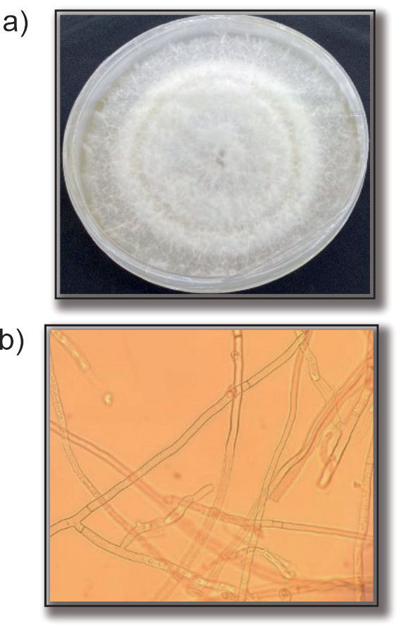
The results of isolating infected plants revealed the acquisition of several isolates. One isolate was selected for each region and assigned symbols (Ry1, Rt1, and Rn1). The three isolates demonstrated growth on Potato Dextrose Agar (PDA), forming colonies with a white color, as shown in Fig. 1. They tended to exhibit a light or dark brown color as they matured. The fungal mycelium was branched, with branching near the terminal end of the fungal hyphae and constriction in the branches. A barrier was formed near the branching zone, and barrel-shaped cells were observed upon microscopic examination, confirming their affiliation with the fungus R. solani, as described by [20, 28].
Testing the Pathogenicity of R. solani Isolates
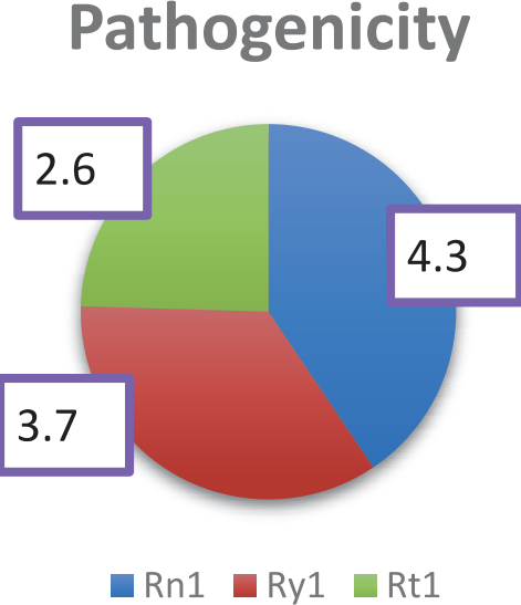
Results of Fig. 2 show that isolate Rn1 had the highest level of pathogenicity (strong), as measured by the length of the discoloration distance on radish seedlings, reaching 4.3, followed by isolate Ry1, which recorded 3.7. In contrast, isolate Rt1 had a medium level of pathogenicity, as described by [21].
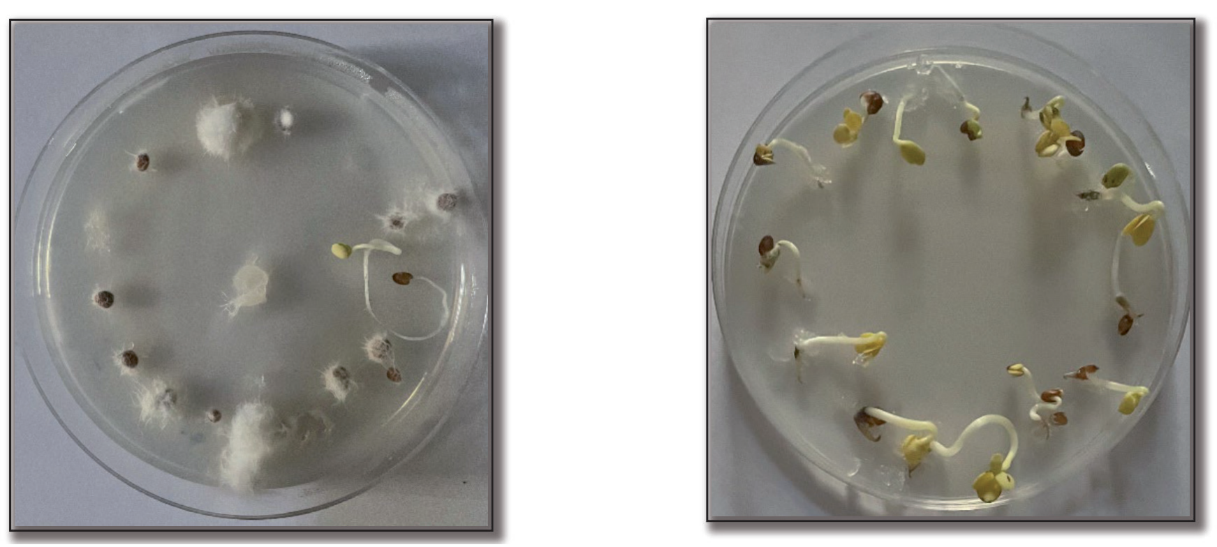
The results, as clarified in Table 2, indicate that all isolates effectively reduced the percentage of seed germination. The Rn1 isolate recorded 6.65, while the Ry1 isolate reached 6.66, and the Rt1 isolate recorded 15.5, compared to the control treatment, which registered 95.3%, as shown in Fig. 3.
The variation in the pathogenicity of isolates may be attributed to their differences in producing enzymes that affect seed germination and variations in the speed and quantity of enzyme production. Consequently, this affects their ability to inhibit or impede seed germination. It may also be attributed to the genetic composition differences among isolates and the geographical variation in the isolated regions of fungal isolates, which influence the survival mechanisms, spread, and occurrence of infections. The ability of R. solani isolates to produce enzymes that break down plant cell walls and cytoplasm, such as cellulase, pectinase, and amylase, causes seed or seedling rot and death [29]. These results align with [1], confirming the variation in the pathogenic ability of isolates isolated from infected potato plants and different regions.
The Molecular Diagnosis of the Most Pathogenic Fungal Isolate
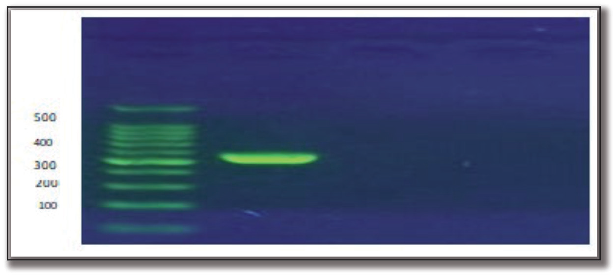
The electrophoresis results of the extracted DNA from the most pathogenic fungal isolate are shown in Fig. 4. On agarose gel electrophoresis, a single band with a molecular weight of 550 bp was observed for the fungal isolate using the ITS1 and ITS4 cloning primers. This result confirmed the ability of these primers to amplify the rDNA nucleic acid for the fungus species R. solani.
Nucleotide Sequence Study
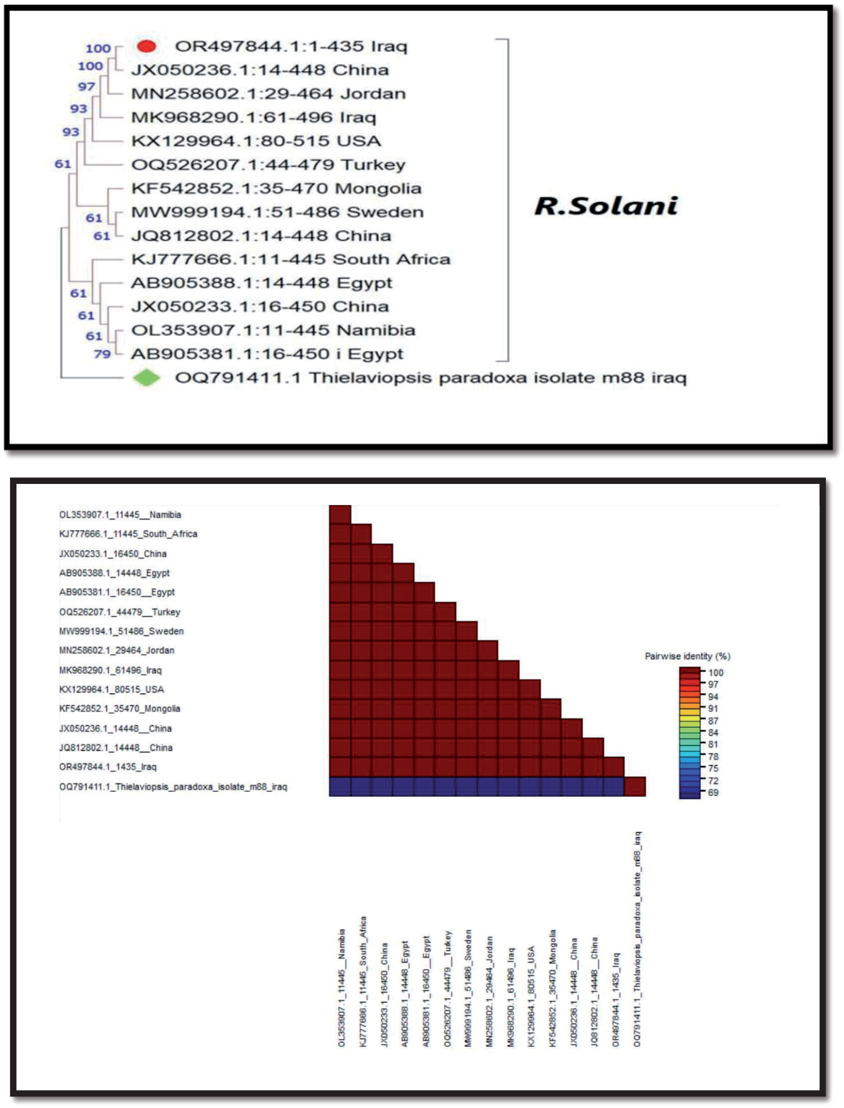
The nucleotide sequencing results revealed that the R. solani fungal isolate exhibited a 100% match with international isolates available in the NCBI global gene bank. The nucleotide sequences were deposited in the World Organization for Gene Banks under accession number OR497844.1, serving as a reference for Iraq, the Middle East, and worldwide. The phylogenetic tree diagram in Fig. 5 illustrates a dendrogram based on molecular sequences of nitrogen base sequences. The tree comprised two clusters, with the outer cluster representing the external isolate Thielaviopsis paradoxa, which is genetically divergent by 69% from all Rhizoctonia fungal isolates. The comparative baseline isolate was present in both clusters, including the Iraqi isolate and isolates from India, Sudan, Mongolia, Turkey, the United States, Jordan, China, and another Iraqi isolate. The second cluster included the South African isolate, the Chinese isolate, the Egyptian isolate, and the Namibian isolate.
The Effect of Aqueous and Alcoholic Extracts of the Calotropis procera Plant and the Fungicide Rizolex on the Fungal Growth of the Pathogenic Fungus R. solani in the Laboratory
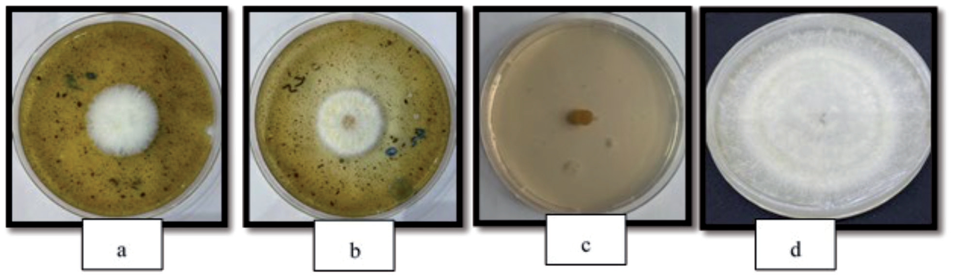
The results in Table 3 demonstrated the effectiveness of both aqueous and alcoholic Calotropis procera extracts in inhibiting R. solani fungus at all concentrations used on PDA medium in the laboratory, compared to the control treatment. The inhibition rate was 64.7% at a concentration of 2 g/100 ml for the aqueous extract, while it reached 62.35% and 56.40% at concentrations of 1% and 0.5%, respectively, showing significant differences between them. The alcoholic extract recorded inhibition rates of 55.29%, 52.32%, and 50.72% at concentrations of 2%, 1%, and 0.5%, respectively, which differed significantly from the pesticide treatment that achieved a 100% inhibition rate, while the control treatment showed a 0% inhibition rate, as shown in Fig. 6.
The effect of the Rizolex pesticide may be attributed to its ability to break down fungal cell walls or its impact on cell reactions, enzymes, and substances secreted by them. The effectiveness of Rizolex in inhibiting the fungus Rhizoctonia solani. The inhibition rates reached 87% at a concentration of 100 mg active substance ml-1 [30].
The effectiveness of Calotropis procera extract in inhibiting fungi is attributed to its secondary metabolites, such as alkaloids, tannins, saponins, and steroids [17]. The acetone extract of Calotropis procera leaves demonstrated efficacy in inhibiting the fungi Penicillium sp., Aspergillus sp., and Aspergillus niger, which are pathogens affecting rice crops, with inhibition percentages reaching 7.1, 7.4, and 7 mm, respectively.
Waheed et al. [31] mentioned that concentrations of 1%, 2.5%, 4%, 5.5%, and 7% achieved inhibition percentages in the growth of the fungus Macrophomina phaseolina, ranging from 15% to 38%. Calotropis procera extract demonstrated effectiveness in inhibiting the growth of Fusarium oxysporum f. sp. Lycopersici, the causative agent of Fusarium wilt in tomatoes, with an 87% inhibition rate at a concentration of 25% [32].
Table 3.
The effectiveness of aqueous and alcoholic brocade extracts in inhibiting the growth of the pathogenic fungus Rhizoctonia solani in the laboratory.
Fig. 6.
The antagonistic potential of the Calotropis procera extract and the pesticide against the fungus Rhizoctonia solani. a) alcoholic Calotropis procera extract, b) aqueous Calotropis procera extract, c) pesticide, d) control.
Mahmood et al. [33] noted the effectiveness of Calotropis procera extract in inhibiting the growth of the fungi Aspergillus flavus and Penicillium notatum in the culture medium. The inhibition percentages were 19.6%, 47.3%, 81.4%, 8.8%, 29.6%, and 59.2% in a sequence for concentrations of 0.5, 1, and 1.5 mg/mL, compared to the control treatment, which recorded a 0% inhibition rate. The inhibition percentages for spore formation were 32.3%, 46.7%, 67.2%, 50.8%, 53.4%, and 70.1% in sequence, reaching 0% for the control treatment.
Santana and Cardoso [16] mentioned that the aqueous extract of Calotropis procera inhibited the growth of the fungi Phomopsis sojae and Sclerotinia sclerotiorum, causative agents of stem dryness, pod blight, and white mold on soybeans, at various concentrations. Effectiveness increased with higher concentrations. The high inhibitory capability of the aqueous Calotropis procera extract may be attributed to its ability to break down its secondary metabolites in water more than in organic solvents, thereby increasing its effectiveness in affecting fungi.
Evaluate the Efficacy of Aqueous Calotropis procera Extract and the Pesticide Rizolex in Controlling Black Scurf Disease on Potatoes Caused by the Fungus Rhizoctonia solani in the Field
The results presented in Table 4 demonstrated the efficacy of aqueous Calotropis procera extract in protecting potato plants from the effects of the fungus R. solani, the causative agent of black scurf disease, and in enhancing growth parameters for the plants. The immersion method of potato tubers in aqueous Calotropis procera extract proved effective in reducing the severity of infection for both the above-ground and below-ground portions affected by the fungus R. solani, with percentages reaching 8.00% and 20.00%, respectively, compared to the conventional treatment method, which recorded infection rates of 28.00% and 48.00% for the above-ground and belowground portions, with significant differences. In contrast, the results indicated the superiority of the pesticide treatment via watering, with infection severity reaching 18.00% for both above-ground and belowground portions. In comparison, it recorded 30.0% and 21.0% for the immersion method with the pesticide. The results highlighted the effectiveness of the extract and the pesticide in reducing infection severity for both above-ground and below-ground portions compared to the control treatment, which reached 80.00% and 86.67%, respectively, with clear significant differences. Thus, the efficacy of the aqueous Calotropis procera extract (via the immersion method) was superior to the pesticide, and its effectiveness can be attributed to the ability of secondary metabolites in Calotropis procera to protect the cell wall from infection by the fungus R. solani.
The results in Table 5 clarified the effectiveness of aqueous Calotropis procera extract through immersion and dusting methods in increasing the plants’ fresh and dry weights. The weights reached 1116.67 g, 72 g, 750 g, and 69 g, respectively, in sequence, compared to the control treatment, which recorded 516.67 g and 44.33 g, respectively. Meanwhile, the pesticide treatment via watering and dusting resulted in weights of 966.67 g, 76 g, 766.67 g, and 71.33 g, respectively. The results indicated all treatments’ superiority in increasing fresh and dry weights compared to the control treatment, with clear significant differences.
An increase in production was observed for all treatments, with the immersion method of aqueous Calotropis procera extract showing the highest productivity at 2116.67 g, followed by the pesticide treatment via watering at 1516.67 g. The aqueous Calotropis procera extract and pesticide treatments via dusting recorded 1150 g each, showing a significant difference from each other and from the control treatment, which registered 616.67 g.
Table 4.
Evaluation of the efficacy of aqueous Calotropis procera extract and the pesticide Rizolex in reducing the severity of infection caused by the fungus Rhizoctonia solani on potato plants.
Table 5.
Evaluation of the efficacy of aqueous Calotropis procera extract and the pesticide Rizolex in growth parameters for the disease caused by the fungus Rhizoctonia solani on potato plants.
Research has indicated the effectiveness of Calotropis procera plant extract in inhibiting fungal growth, promoting seed germination, improving growth parameters, and increasing plant productivity. Olaitan et al. [17] mentioned that extracts from the leaves and latex of Calotropis procera inhibited the growth of fungi such as Cuvularia lunata, Alternaria alternata, Rhizoctonia solani, Fusarium solani, Penicillium chrysogenum, Aspergillus niger, A. flavus, A. terrus, A. fumigatus, and Rhizopus sp. transmitted by soybean, sunflower, mustard, and peanut seeds. Ahmed et al. [34] reported that Calotropis procera plant extract demonstrated high efficacy in reducing fungi transmitted by barley seeds and increasing seed germination rates at a concentration of 20%. Rani et al. [35] highlighted the efficacy of Calotropis procera in protecting plants from various fungi such as Phaeoramularia calotropidis, Curvularia hawaiiensis, Guignardia bidwellii, Alternaria alternata, Cochliobolus hawaiiensis, Aspergillus spp., Mucor circinelloides, Fusarium spp., Chaetomium spp., and Penicillium spp. Mahmood et al. [33] indicated that Calotropis procera extract at concentrations of 1 and 1.5 ml increased wheat germination percentage, reaching 80% and 93.3%, respectively, compared to the control treatment, which was 73.3%. Results from the plastic house experiment showed that Calotropis procera plant extract significantly improved the vegetative growth of tomato plants, reaching 20.08 grams compared to the control treatment, which recorded 10.20 grams [15]. Almaghasla et al. [30] reported that the fungicide Rizolex demonstrated high efficacy against the fungus Rhizoctonia solani in the greenhouse. The percentage of healthy seedlings reached 73.3% when treated with the fungicide before planting, compared to other pesticide treatments. Additionally, Rizolex reduced the number of spots to 0.22 spots/leaf caused by the fungus Alternaria alternata. The effectiveness of the pesticide through watering may be attributed to its persistence in the soil and its greater influence on the pathogenic agent. Even though resistance in plants against pathogens is the most stable strategy to avoid losses in agriculture [36], plant extracts are rising as a promising solution to reduce the dependence on excessive use and misuse of pesticides in agriculture to reduce the damage of pathogens [37, 38].

